BiteSized Immunology: Cells
Cells T CD8+
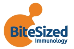
CD8+ (cytotoxic) T cells, like CD4+ Helper T cells, are generated in the thymus and express the T-cell receptor. However, rather than the CD4 molecule, cytotoxic T cells express a dimeric co-receptor, CD8, usually composed of one CD8α and one CD8β chain. CD8+ T cells recognise peptides presented by MHC Class I molecules, found on all nucleated cells. The CD8 heterodimer binds to a conserved portion (the α3 region) of MHC Class I during T cell/antigen presenting cell interactions (see Figure 1).
CD8+ T cells (often called cytotoxic T lymphocytes, or CTLs) are very important for immune defence against intracellular pathogens, including viruses and bacteria, and for tumour surveillance. When a CD8+ T cell recognises its antigen and becomes activated, it has three major mechanisms to kill infected or malignant cells. The first is secretion of cytokines, primarily TNF-α and IFN-γ, which have anti-tumour and anti-viral microbial effects.

The second major function is the production and release of cytotoxic granules. These granules, also found in NK cells, contain two families of proteins, perforin, and granzymes. Perforin forms a pore in the membrane of the target cell, similar to the membrane attack complex of complement. This pore allows the granzymes also contained in the cytotoxic granules to enter the infected or malignant cell. Granzymes are serine proteases which cleave the proteins inside the cell, shutting down the production of viral proteins and ultimately resulting in apoptosis of the target cell.
The cytotoxic granules are released only in the direction of the target cell, aligned along the immune synapse, to avoid non-specific bystander damage to healthy surrounding tissue (see Figure 1). CD8+ T cells are able to release their granules, kill an infected cell, then move to a new target and kill again, often referred to as serial killing.
The third major function of CD8+ T cell destruction of infected cells is via Fas/FasL interactions. Activated CD8+ T cells express FasL on the cell surface, which binds to its receptor, Fas, on the surface of the target cell. This binding causes the Fas molecules on the surface of the target cell to trimerise, which pulls together signalling molecules. These signalling molecules result in the activation of the caspase cascade, which also results in apoptosis of the target cell. Because CD8+ T cells can express both molecules, Fas/FasL interactions are a mechanism by which CD8+ T cells can kill each other, called fratricide, to eliminate immune effector cells during the contraction phase at the end of an immune response.
In addition to their critical role in immune defense against viruses, intracellular bacteria, and tumours, CD8+ T cells can also contribute to an excessive immune response that leads to immunopathology, or immune-mediated damage.
© The copyright for this work resides with the author